Your eyes are said to be one of the most complex organs in your body. The intricate way in which they work will be explained below.
They work like a camera; or, rather the camera was made to work like them. The simplest explanation involves the detection of light and conversion into electrochemical impulses in neurons.
The light focuses on your retina and the retina reacts like the film in a camera by sending a record of it by means of your optic nerve to your brain.
The inner workings of your eyes are a superlative design involving many complex functions and connections that no man has been able to duplicate. To this date, no one has been able to come remotely close to copying it.
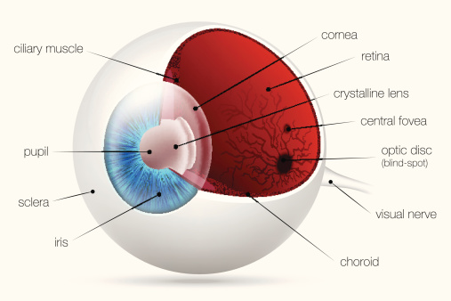
Some of the main parts of the human eye:
1. Iris
2. Lens
3. Retina
4. Sclera
5. Macula
6. Cornea
7. Optic Nerve
The Iris: This is the colored circle of tissue that is behind the cornea. It controls the diameter and size of your pupil and the amount of light that reaches the retina. The color of the iris is different because of our genetic makeup. In the center of your iris is the circular pupil.
The Lens: This is directly behind your iris. It is transparent and flexible and its shape is controlled by muscle fibers. The lens makes the final focus adjustments of light rays that come into your eye so that they are sharply focused on the retina.
The Retina: This is the innermost layer of the wall of your eye. It consists of light sensitive cells that absorb light rays and change them into electrical signals that are passed to the brain and then interpreted as visual images.
The Sclera: This is known as the white part of the eye. It is opaque and fibrous. It acts as the protective layer of your eye.
The Macula: This is commonly known as the yellow “spot” on your eye. It is the oval-shaped and highly pigmented yellow spot near the center of the retina. It is responsible for absorbing excess blue and ultraviolet light that comes into your eye. It acts like a natural “sunblock” for this area of the retina. The yellow color comes from what are called lutein and zeaxanthin, which are yellow xanthophyll carotenoids, which come from our diet.
The Cornea: This is the transparent part of your eye that covers the iris, pupil and the anterior chamber. Working with the lens to refract light, it contributes to the ability of the eye to focus. The cornea causes involuntary reflexes to close your eye because of itr s unmyelinated nerve endings. It has no blood vessels.
Optic Nerve: This is also known as your cranial nerve 2 because it transmits visual information from your retina to your brain. It is the second of twelve paired cranial nerves and is considered as part of your central nervous system. Your optic nere has three meningeal layers. It is composed of retinal ganglion cell axons and support cells.
The complexity of our eyes goes much deeper than this explanation here, and its detail of how they work could take much time to study. Many that have studied the human eye have said it certainly is a marvel of a Supreme Designer.